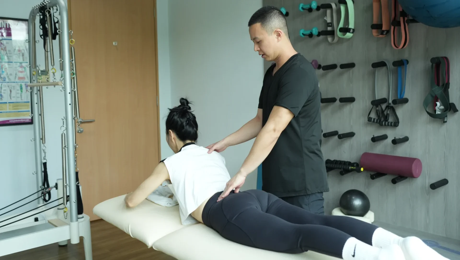
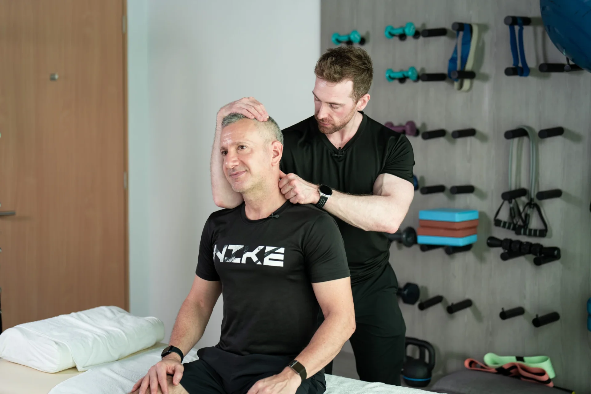
Few musculoskeletal diagnoses generate more fear and more confusion than “slipped disc.” The label itself is misleading, the imaging findings are widely misinterpreted, and the treatment pathway that most patients end up on is often not the one the evidence supports. For a condition that affects a substantial proportion of the working population in Singapore at some point in their lives, the gap between what most people believe about slipped discs and what the clinical evidence actually shows is one of the most consequential in musculoskeletal medicine.
This piece is a research-grounded guide to what a slipped disc actually is, what the symptoms look like, how recovery typically unfolds, and when physiotherapy is the right first step. It is calibrated to the specific presentation that adults in Singapore, particularly those with desk-based occupations and prolonged sitting patterns, most commonly experience.
The framing this piece sits within is the Two-Layer Problem covered in depth in the TSquared Physio Osteopathy and Physiotherapy library: persistent back pain and disc-related presentations almost always involve both a structural layer (the disc pathology itself and the joint restrictions around it) and a functional layer (the movement patterns, weakness, and compensatory guarding that either resolve or entrench the presentation). Understanding both is the difference between recovery and recurrence.
What a Slipped Disc Actually Is

The specific type shapes the treatment. The label does not.
The term “slipped disc” is anatomically inaccurate. Intervertebral discs do not slip out of position. What actually happens is that the soft inner core of the disc (the nucleus pulposus) pushes against or through the tougher outer ring (the annulus fibrosus), producing what clinicians more accurately call a disc bulge, disc protrusion, disc herniation, or disc extrusion depending on the specific pattern and severity.
The clinical significance of the distinction is that “slipped disc” implies a mechanical dislocation that requires manual repositioning, when the reality is a tissue-level change in the disc structure that responds to a specific set of clinical interventions. The language shapes the expectation, and the expectation shapes the treatment path.
The Grades of Disc Pathology
Disc pathology exists on a spectrum, and the language matters for treatment decisions:
Disc bulge: the disc extends beyond its normal boundary but the outer ring remains intact. This is the most common and least severe pattern. Disc bulges are present in a substantial proportion of asymptomatic adults and often do not require treatment when they are not producing symptoms.
Disc protrusion: the inner core pushes further against the outer ring, producing a more focal deformity of the disc shape. Protrusions may or may not produce nerve root symptoms depending on their location and size.
Disc herniation (extrusion): the inner core breaks through the outer ring but remains connected to the disc. This more commonly produces nerve root symptoms and is the pattern most people associate with the term “slipped disc.”
Disc sequestration: a fragment of the inner core detaches from the disc entirely. This is the most severe pattern and more commonly requires surgical evaluation.
The Imaging Interpretation Problem
One of the most consistent findings in the spinal imaging literature is that disc abnormalities are common in asymptomatic adults. Brinjikji and colleagues, in a systematic review of imaging findings in asymptomatic populations, found that disc degeneration was present in 37 percent of asymptomatic 20-year-olds and 96 percent of asymptomatic 80-year-olds. Disc bulges were present in 30 percent of asymptomatic 20-year-olds and 84 percent of asymptomatic 80-year-olds (Brinjikji et al., 2015).
The clinical significance is that an MRI showing a disc bulge or protrusion does not, on its own, explain the presence or severity of a patient’s back pain. Imaging findings must be interpreted in the context of the clinical presentation, the symptom pattern, and the physical examination, rather than used as the primary basis for treatment decisions. This is the Clean Report Problem in reverse: imaging that produces a finding is not always a finding that requires treatment.
Symptoms of a Slipped Disc

The symptom pattern of a symptomatic disc herniation varies depending on the location and severity of the herniation, and on which nerve structures are affected. The most common presentations fall into recognisable clinical patterns.
Lower Back Pain
Localised lower back pain is the most common symptom of lumbar disc pathology. The pain is typically deep, aching, and worsened by specific movements: bending forward, sitting for extended periods, lifting, or twisting. Extension movements (bending backward) may relieve or worsen the pain depending on the specific pattern of the disc pathology.
Morning stiffness that improves with movement, worsening pain across a prolonged sitting day, and difficulty finding a comfortable sleeping position are common accompaniments.
Radicular Pain (Sciatica)
When a disc herniation compresses or irritates a nerve root, the pain pattern changes. Instead of remaining localised in the lower back, the pain radiates down the leg, following the distribution of the affected nerve. This is commonly called sciatica when it involves the sciatic nerve, though the term is often used loosely to describe any leg pain of spinal origin.
The classic sciatic distribution runs from the lower back, through the buttock, down the back of the thigh, into the calf, and sometimes into the foot. The specific pattern varies depending on which nerve root is affected. The pain is often described as sharp, burning, or electric, and may be worse than the back pain itself.
Neurological Symptoms
More significant nerve root compression can produce neurological symptoms beyond pain: numbness or altered sensation in the leg or foot, weakness in specific muscle groups (foot drop being the most recognised pattern), and reduced or absent reflexes on clinical examination.
These are more concerning findings that warrant thorough clinical assessment and, in some cases, imaging and specialist referral.
Red Flag Symptoms
A small subset of symptoms warrant urgent medical assessment rather than routine physiotherapy referral. These include:
- Loss of bladder or bowel control
- Numbness in the saddle region (perineum, inner thighs, buttocks)
- Progressive weakness in both legs
- Severe and worsening pain despite rest
- Fever, unexplained weight loss, or history of cancer with new back pain
These findings can indicate cauda equina syndrome or other conditions requiring immediate medical attention. Any patient with these symptoms should present to a hospital emergency department rather than book a physiotherapy appointment.
How Recovery Actually Unfolds
The recovery trajectory for symptomatic disc herniations is often better than the diagnosis leads patients to expect. This is one of the most under-communicated aspects of the condition.
The Natural History
Systematic reviews of the natural history of lumbar disc herniation have consistently demonstrated that a significant proportion of symptomatic disc herniations improve spontaneously over weeks to months. The herniated disc material itself often reduces in size over time through a process called resorption, mediated by inflammatory and immune responses that gradually break down the extruded disc tissue (Chiu et al., 2015).
Approximately two-thirds of patients with acute sciatica from disc herniation experience meaningful improvement within six to twelve weeks with conservative management. This does not mean the condition resolves without intervention. It means that with appropriate physiotherapy, load management, and time, the trajectory is often toward recovery rather than deterioration.
The Acute Phase
The first two to six weeks after symptom onset is the acute phase. Pain is typically most severe during this period, and the priority is symptom management, appropriate load modification, and early gentle movement rather than aggressive intervention.
Bed rest, once widely recommended, is no longer supported by the evidence. Prolonged bed rest actually slows recovery by deconditioning the supporting musculature and reducing the mobility the spine requires to heal. Current guidelines recommend maintaining activity within tolerance and avoiding positions and movements that clearly aggravate symptoms.
The Subacute Phase
Between six and twelve weeks, most patients experience meaningful reduction in acute symptoms and can begin more active rehabilitation. This is the phase where physiotherapy earns its place most clearly: progressive loading, movement re-education, addressing the compensatory patterns that developed during the acute phase, and building the functional capacity that prevents recurrence.
The Pain Loop, covered in depth in the TSquared Physio Chronic Back Pain library, is directly relevant here. Protective guarding that developed during the acute phase, if not deliberately addressed, becomes entrenched dysfunction that outlasts the disc healing itself. This is one of the most consistent reasons why patients experience persistent back pain long after the disc pathology has resolved.
The Longer-Term Picture
Beyond twelve weeks, the majority of patients have returned to normal or near-normal function with appropriate management. A smaller subset experiences persistent symptoms that require longer-term management. For these patients, the clinical question shifts from “will this resolve” to “how do we manage this sustainably,” and the physiotherapy approach adjusts accordingly.
Recurrence is a real consideration. The physical loading patterns, movement habits, and postural adaptations that contributed to the original disc presentation are often still present after acute recovery. Without addressing these underlying factors, the recurrence rate is substantial.
When Physiotherapy Is the Right First Step

For the majority of symptomatic disc herniations without red flag symptoms, physiotherapy is the appropriate first-line intervention. The evidence supporting this position is well-established.
Clinical guidelines from major professional bodies, including the American College of Physicians, the National Institute for Health and Care Excellence (NICE) in the UK, and international pain management consensus statements, consistently recommend conservative management (physiotherapy, exercise, appropriate pain management) as the first-line approach for non-specific lower back pain and for most lumbar disc herniations without progressive neurological deficit (Qaseem et al., 2017).
What Physiotherapy Actually Does
Physiotherapy for a symptomatic disc herniation is not a single intervention. It is a progressive clinical programme that adjusts to the stage of recovery and the specific presentation.
Acute phase interventions focus on symptom management, appropriate load modification, and early gentle movement. Manual therapy, specific mobility work, and postural adjustments that reduce mechanical load on the affected segment are the priorities.
Subacute phase interventions introduce progressive loading, movement re-education, and addressing the compensatory patterns that developed during the acute phase. Specific exercises targeting the deep core stabilisers, the gluteal complex, and the movement patterns most likely to prevent recurrence become the centrepiece of the programme.
Return-to-function interventions rebuild the functional capacity required for the patient’s specific work, sport, and daily life demands. This is where the programme moves from managing the acute presentation to building the resilience that prevents recurrence.
The Two-Layer Response
A symptomatic disc herniation is a textbook expression of the Two-Layer Problem. The structural layer includes the disc pathology itself, the joint restrictions that developed around it, and the tissue tension patterns that accumulate through protective guarding. The functional layer includes the movement patterns, muscle weakness, and neuromuscular compensation that either resolve or entrench the presentation.
Addressing only one layer produces incomplete recovery. Physiotherapy that focuses exclusively on symptom management without rebuilding functional capacity leaves the patient vulnerable to recurrence. Manual therapy without progressive loading resolves the acute presentation but does not build the resilience the spine requires. At TSquared Physio, both layers are addressed within a single coordinated programme, with osteopathy and physiotherapy working as integrated modalities rather than separate services.
When to Consider Surgery
Surgery for lumbar disc herniation is appropriate for a specific and relatively narrow set of clinical presentations: progressive neurological deficit, cauda equina syndrome, severe and persistent symptoms unresponsive to conservative management after an adequate trial (typically six to twelve weeks minimum), or specific occupational or functional requirements that make longer-term conservative management impractical.
For the majority of symptomatic disc herniations, surgery is not the appropriate first-line intervention. Systematic reviews comparing surgical and conservative management for lumbar disc herniation have generally found that surgical outcomes may be somewhat better in the short term for pain relief and return to work, but that long-term outcomes converge, and that most patients who choose conservative management achieve satisfactory outcomes without surgery (Weinstein et al., 2008).
The decision to pursue surgery should be made in consultation with a spinal specialist, based on the specific clinical presentation, the response to an adequate trial of conservative management, and the patient’s specific goals and circumstances.
What Slows Recovery
Several factors are associated with slower recovery from lumbar disc herniation, and understanding them helps calibrate expectations and identify the interventions most likely to make a difference.
Prolonged Sitting and the Sitting Debt
Sustained sitting increases intradiscal pressure and reduces the movement and load variability the disc requires to remain healthy. For Singapore-based professionals whose working day involves 8 to 12 hours of sitting, the postural and mechanical load on the lumbar spine is substantial and directly relevant to both the development and the recovery from disc pathology.
This is the Sitting Debt covered in depth in the TSquared Physio library. The structural consequences of prolonged sitting on the lumbo-pelvic-hip complex are not a separate issue from disc recovery. They are directly connected. A disc recovery programme that does not address the sitting pattern is treating the symptom while the underlying mechanical load continues.
Fear-Avoidance and Movement Patterns
Fear of pain and fear of re-injury are among the most consistent predictors of poor recovery from lumbar disc herniation. Patients who become fearful of movement, who avoid activities that produced pain in the past, and who develop protective guarding patterns often experience longer recovery timelines than patients who remain appropriately active within their tolerance.
This is one of the reasons why appropriate education, graduated exposure to movement, and confident clinical communication are core components of physiotherapy for disc pathology. The psychological dimension is not separate from the physical recovery. It shapes it directly.
Underlying Deconditioning
Patients whose pre-injury conditioning is limited (weak core stabilisers, poor gluteal activation, restricted hip mobility, limited thoracic mobility) typically experience longer and more complex recoveries than patients with better baseline conditioning. The physiotherapy programme addresses these underlying factors directly, but they cannot be resolved in the acute phase.
Systemic Factors
Chronic inflammation, poor sleep, metabolic dysfunction, and hormonal factors all influence tissue healing and recovery speed. For patients whose recovery is slower than expected despite appropriate physiotherapy, systemic factors may be contributing. The Recovery Illusion covered in the TSquared Health library is directly relevant here: subjective assessment of recovery does not always match the biological picture, and biomarker assessment through TSquared Health can identify systemic contributors that are shaping the recovery trajectory.
What to Expect from a TSquared Physio Assessment
The initial physiotherapy assessment for a suspected disc herniation is thorough. It includes a detailed clinical history mapping the onset, progression, and current symptom pattern; a movement and postural assessment identifying the specific patterns aggravating and relieving symptoms; a neurological screening for nerve root involvement; and where indicated, coordination with a spinal specialist or referral for imaging.
The assessment produces a clinical picture that guides the physiotherapy programme. The programme is calibrated to the specific presentation, the stage of recovery, and the patient’s goals. Progression is guided by clinical response rather than a generic timeline.
For patients whose presentation involves complex mechanical or systemic contributors, coordination with adjacent clinical services is built into the model. Osteopathy is integrated where the structural layer of the Two-Layer Problem warrants it. TSquared Health biomarker assessment is available where systemic contributors are affecting recovery. TSquared Lab strength programming is coordinated with the physiotherapy findings for patients whose return-to-function includes structured training.
Recovery Timelines and Return to Activity
The specific timeline for recovery from a symptomatic disc herniation varies significantly between patients. The following framework provides realistic expectations for the majority of presentations.
Week 0 to 2 (acute phase): Priority on symptom management, appropriate load modification, and early gentle movement. Return to normal daily activities within tolerance. Avoid positions and movements that clearly aggravate symptoms. Bed rest is not recommended.
Week 2 to 6 (early subacute phase): Meaningful reduction in acute symptoms in most patients. Introduction of specific mobility work and gentle progressive loading. Return to light work activities where possible.
Week 6 to 12 (subacute phase): Progressive loading, movement re-education, and addressing compensatory patterns. Return to most work and daily activities. Careful reintroduction of exercise activities. This is the phase where physiotherapy earns its place most clearly.
Week 12 and beyond: Return to full function for most patients. Continued rehabilitation for those with more complex presentations. Focus shifts to building the resilience that prevents recurrence. Return to sport and higher-impact activities becomes appropriate for most patients.
Return to specific activities depends on the demands of the activity and the patient’s current functional capacity. Return to running, resistance training, contact sports, and other high-demand activities should be guided by clinical assessment rather than time alone.
Physiotherapy at TSquared Physio for Disc Presentations

At TSquared Physio, the assessment and treatment of lumbar disc presentations sits within the integrated clinical model that addresses both the structural and functional layers of the Two-Layer Problem. Every patient begins with an extended clinical assessment that maps the specific presentation, the contributing factors, and the recovery goals.
The programme built from that assessment is calibrated to the stage of recovery, the specific mechanical presentation, and the patient’s functional demands. Osteopathy is integrated where the structural layer warrants it. Where the patient’s recovery involves a return to structured training, coordination with TSquared Lab ensures the loading programme progresses within the parameters established by the physiotherapy assessment. Where systemic factors are contributing to slower-than-expected recovery, TSquared Health biomarker assessment provides the physician-led diagnostic picture that supports the clinical response.
For Singapore-based professionals whose working pattern involves prolonged sitting, the Sitting Debt component of the recovery programme becomes particularly relevant. The mechanical load of the working day is not separate from the recovery. It shapes it directly.
The majority of symptomatic disc herniations recover well with appropriate physiotherapy. The programme is calibrated to what the specific presentation requires, and the recovery is guided by clinical response rather than fear or generic timelines.
Book an appointment with TSquared Physio at tsquaredphysio.com or contact the clinical tea
FAQs
What is the difference between a slipped disc, a bulging disc, and a herniated disc?
The terms describe different points on the same spectrum of disc pathology. A bulging disc extends beyond its normal boundary while the outer ring remains intact. A protrusion involves a more focal deformity of the disc shape. A herniation involves the inner core breaking through the outer ring but remaining connected to the disc. “Slipped disc” is a colloquial term that is not anatomically accurate: discs do not slip out of position. The clinical significance of the specific type depends more on whether nerve structures are affected and how the presentation responds to conservative management than on the label itself.
Do I need an MRI if I think I have a slipped disc?
Not always. MRI is appropriate when the clinical presentation includes progressive neurological deficit, red flag symptoms suggesting more serious pathology, or when the patient has not responded to an adequate trial of conservative management. For the majority of symptomatic disc presentations without red flags, imaging is not required to guide initial physiotherapy, and premature imaging can actually complicate the clinical picture because disc abnormalities are so common in asymptomatic adults (Brinjikji et al., 2015). A clinical assessment determines whether imaging is warranted in a specific case.
How long does a slipped disc take to heal?
The natural history of symptomatic disc herniation is generally favourable. Approximately two-thirds of patients with acute sciatica from disc herniation experience meaningful improvement within six to twelve weeks with appropriate conservative management. The herniated disc material itself often reduces in size over time through resorption. The specific timeline varies significantly between patients depending on the presentation, the contributing factors, and the response to intervention.
Can I exercise with a slipped disc?
Yes, in most cases, and appropriate exercise is part of the recovery. Bed rest is no longer recommended for lumbar disc herniation. Current guidelines recommend maintaining activity within tolerance and avoiding positions and movements that clearly aggravate symptoms. The specific exercises appropriate for a given presentation depend on the stage of recovery, the specific symptom pattern, and the response to different movement patterns. A physiotherapy assessment provides the framework for calibrating exercise appropriately.
Do I need surgery for a slipped disc?
For the majority of symptomatic disc herniations, surgery is not the appropriate first-line intervention. Systematic reviews comparing surgical and conservative management have generally found that long-term outcomes converge, and that most patients achieve satisfactory outcomes without surgery (Weinstein et al., 2008). Surgery is appropriate for progressive neurological deficit, cauda equina syndrome, severe and persistent symptoms unresponsive to conservative management after an adequate trial, or specific occupational requirements. The decision should be made in consultation with a spinal specialist based on the specific clinical picture.
What are the red flags I should watch for?
Any of the following symptoms warrant urgent medical assessment rather than routine physiotherapy: loss of bladder or bowel control, numbness in the saddle region (perineum, inner thighs, buttocks), progressive weakness in both legs, severe and worsening pain despite rest, fever, unexplained weight loss, or history of cancer with new back pain. These findings can indicate cauda equina syndrome or other conditions requiring immediate medical attention. Patients with these symptoms should present to a hospital emergency department.
Will my slipped disc come back?
Recurrence is a real consideration. The physical loading patterns, movement habits, and postural adaptations that contributed to the original disc presentation are often still present after acute recovery. Without addressing these underlying factors, the recurrence rate is substantial. This is one of the reasons why the functional layer of the Two-Layer Problem, building the strength, movement quality, and postural resilience that prevents recurrence, is as important as managing the acute symptoms.
Can prolonged sitting cause a slipped disc?
Prolonged sitting increases intradiscal pressure and reduces the movement and load variability the disc requires to remain healthy. For Singapore-based professionals whose working day involves 8 to 12 hours of sitting, the postural and mechanical load on the lumbar spine is substantial and is a contributing factor to disc pathology. Prolonged sitting alone does not typically cause an acute disc herniation, but it contributes to the underlying conditions in which disc pathology develops and recurs. The Sitting Debt covered in depth in the TSquared Physio library addresses these consequences directly.